
Exercise 14 gross anatomy of the brain and cranial nerves – Embarking on Exercise 14: Gross Anatomy of the Brain and Cranial Nerves, we delve into the intricacies of the central nervous system, exploring the brain’s major regions and their functions, while unraveling the pathways and functions of the 12 cranial nerves.
This exploration provides a foundation for comprehending neurological disorders and their diagnosis and management.
Delving deeper, we examine the microscopic structure of the brain and cranial nerves, deciphering the cellular components and their roles. The developmental journey of these structures is charted, tracing their embryonic origins through various stages, highlighting potential developmental disruptions. Comparative anatomy unveils the brain and cranial nerve variations across species, shedding light on evolutionary relationships and implications for understanding human brain function.
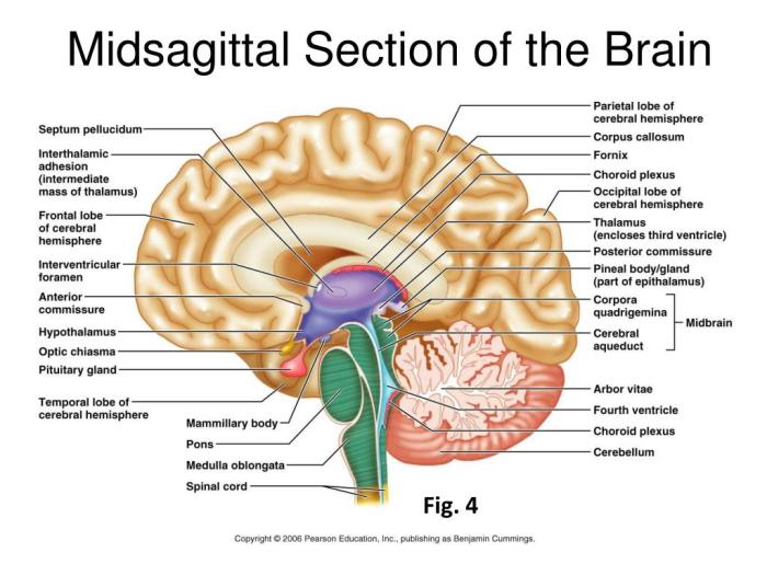
Gross Anatomy of the Brain
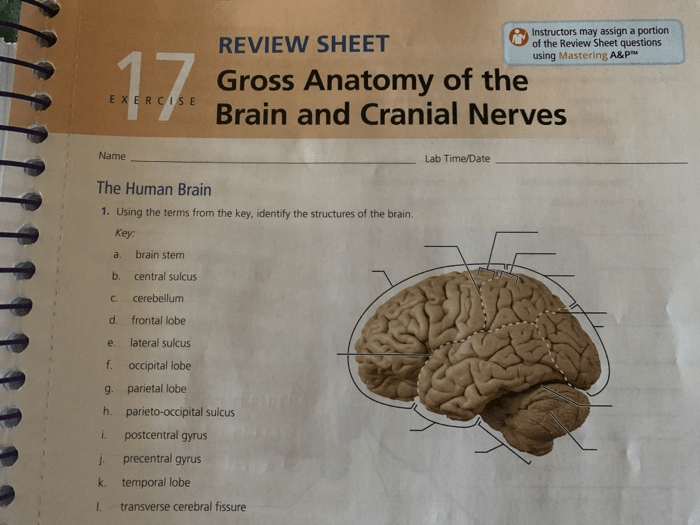
The brain is the control center of the nervous system, responsible for regulating vital functions, processing sensory information, and generating thoughts and emotions. It is composed of three major regions: the forebrain, midbrain, and hindbrain.
Forebrain
The forebrain includes the cerebrum, diencephalon, and basal ganglia.
- Cerebrum:The largest part of the brain, responsible for higher-level functions such as cognition, memory, and language.
- Diencephalon:Contains the thalamus and hypothalamus, which relay sensory information and regulate endocrine function.
- Basal ganglia:Involved in motor control and coordination.
Midbrain
The midbrain connects the forebrain to the hindbrain and contains structures involved in motor control, auditory processing, and visual reflexes.
Hindbrain, Exercise 14 gross anatomy of the brain and cranial nerves
The hindbrain includes the cerebellum, pons, and medulla oblongata.
- Cerebellum:Coordinates movement and balance.
- Pons:Relays sensory information and motor commands.
- Medulla oblongata:Controls vital functions such as breathing, heart rate, and blood pressure.
Cranial Nerves: Exercise 14 Gross Anatomy Of The Brain And Cranial Nerves

The cranial nerves are 12 pairs of nerves that originate from the brain and extend to various parts of the head, neck, and trunk.
Origin, Pathway, and Distribution
Each cranial nerve has a specific origin, pathway, and distribution, innervating specific structures and performing distinct functions.
Functions
Cranial nerves are responsible for a wide range of functions, including:
- Sensory perception (e.g., vision, hearing, smell)
- Motor control (e.g., facial movements, tongue movement)
- Autonomic functions (e.g., heart rate regulation, digestion)
Clinical Significance
A thorough understanding of brain and cranial nerve anatomy is crucial for diagnosing and treating neurological disorders.
Neurological Disorders
Common neurological disorders that affect the brain and cranial nerves include:
- Stroke
- Alzheimer’s disease
- Parkinson’s disease
Imaging Techniques
Imaging techniques such as MRI and CT scans play a vital role in diagnosing and managing these disorders by providing detailed images of the brain and cranial nerves.
Detailed FAQs
What are the major regions of the brain?
The major regions of the brain include the cerebrum, cerebellum, brainstem, and diencephalon.
What is the function of the vagus nerve?
The vagus nerve is responsible for regulating various bodily functions, including digestion, heart rate, and respiration.
How can imaging techniques assist in diagnosing neurological disorders?
Imaging techniques such as MRI and CT scans provide detailed images of the brain and cranial nerves, aiding in the diagnosis of neurological disorders.
